Calibrating 3D Scanner in the Coordinate System of Optical Tracker for Image-To-Patient Registration
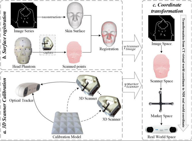
Abstract—Three-dimensional scanners have been widely applied in image-guided surgery (IGS) given its potential to solve the image-to-patient registration problem. How to perform a reliable calibration between a 3D scanner and an external tracker is especially important for these applications. This study proposes a novel method for calibrating the extrinsic parameters of a 3D scanner in the coordinate system of an optical tracker. We bound an optical marker to a 3D scanner and designed a specified 3D benchmark for calibration. We then proposed a two-step calibration method based on the pointset registration technique and nonlinear optimization algorithm to obtain the extrinsic matrix of the 3D scanner. We applied repeat scan registration error (RSRE) as the cost function in the optimization process. Subsequently, we evaluated the performance of the proposed method on a recaptured verification dataset through RSRE and Chamfer distance (CD). In comparison with the calibration method based on 2D checkerboard, the proposed method achieved a lower RSRE (1.73 mm vs. 2.10, 1.94, and 1.83 mm) and CD (2.83 mm vs. 3.98, 3.46, and 3.17 mm). We also constructed a surgical navigation system to further explore the application of the tracked 3D scanner in image-to-patient registration. We conducted a phantom study to verify the accuracy of the proposed method and analyze the relationship between the calibration accuracy and the target registration error (TRE). The proposed scanner-based image-to-patient registration method was also compared with the fiducial-based method, and TRE and operation time (OT) were used to evaluate the registration results. The proposed registration method achieved an improved registration efficiency (50.72 ± 6.04 vs. 212.97 ± 15.91 s in the head phantom study). Although the TRE of the proposed registration method met the clinical requirements, its accuracy was lower than that of the fiducial-based registration method (1.79 ± 0.17 mm vs. 0.92 ± 0.16 mm in the head phantom study). We summarized and analyzed the limitations of the scanner-based image-to-patient registration method and discussed its possible development.

