Improved U-Net for guidewire tip segmentation in X-ray fluoroscopy images
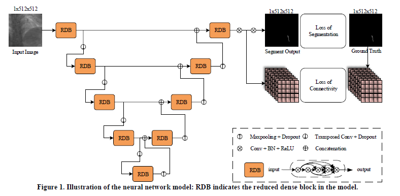
Abstract—In percutaneous coronary intervention (PCI), physicians use a guidewire tip to implant stents in vessels with stenosis. Given the small scale and low signal-to-noise ratio of guidewire tips in X-ray fluoroscopy images, physicians experience difficulty in recognizing and locating the tip. The automatic segmentation of the guidewire tip can ease navigation when the physicians implant stents for PCI. In this paper, we propose an end-to-end convolutional neural network-based method for guidewire tip segmentation. The network framework is derived from U-Net, and two specific designs involving reduced dense block and connectivity supervision are embedded in the framework to improve the accuracy and robustness of guidewire tip segmentation. Experiments are performed on clinical data. The proposed method achieves mean sensitivity, F1-score, Jaccard index, Hausdorff distance of 92.95%, 91.35%, 84.14%, and 0.531 mm on testing data, respectively. In addition, the segmentation time is 0.02 s/frame, which can satisfy the requirements for clinical intra-practice.

